The circulatory system
Cloze Test Worksheet
Kindly Shared By:
Date Shared: 9 June 2022
Worksheet Type:
Tags Describing Content or Audience:
Worksheet Instructions:
None provided.
Target Language or Knowledge:
Arteries
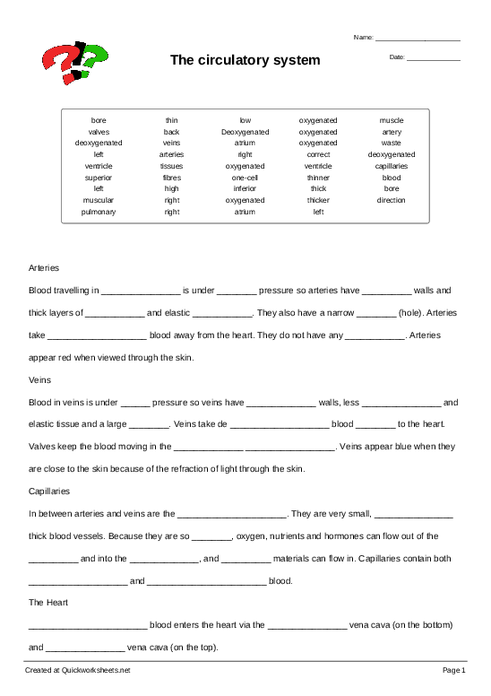
Blood travelling in arteries is under high pressure so arteries have thick walls and thick layers of muscle and elastic fibres. They also have a narrow bore (hole). Arteries take oxygenated blood away from the heart. They do not have any valves. Arteries appear red when viewed through the skin.
Veins
Blood in veins is under low pressure so veins have thinner walls, less muscular and elastic tissue and a large bore. Veins take de oxygenated blood back to the heart. Valves keep the blood moving in the correct direction. Veins appear blue when they are close to the skin because of the refraction of light through the skin.
Capillaries
In between arteries and veins are the capillaries. They are very small, one-cell thick blood vessels. Because they are so thin, oxygen, nutrients and hormones can flow out of the blood and into the tissues, and waste materials can flow in. Capillaries contain both oxygenated and deoxygenated blood.
The Heart
Deoxygenated blood enters the heart via the inferior vena cava (on the bottom) and superior vena cava (on the top).
These blood vessels empty into the right atrium. From here blood flows into the right ventricle, before being pumped to the lungs via the pulmonary artery. The word pulmonary comes from the Latin word pulmonem - meaning “of the lungs”. The pulmonary artery is the only artery in the body to carry deoxygenated blood. The
pulmonary artery divides in two shortly after leaving the heart, in order to deliver blood to the left and right lungs. Upon reaching the lungs, the blood becomes oxygenated and returns to the heart via the pulmonary veins. The pulmonary veins are the only veins in the body to carry oxygenated blood. The pulmonary veins enter into the left atrium. Blood flows from here into the left ventricle. The left ventricle then contracts and sends oxygenated blood to the body via the aorta. The two sides of the heart are divided by a wall called the septum.
The muscular walls of the left ventricle are thicker than the walls of the right ventricle. This is because the right ventricle only needs to pump blood to the lungs, whereas the left ventricle needs to pump blood to the entire body.
arteries high thick muscle fibres bore oxygenated valves low thinner muscular bore oxygenated back correct direction capillaries one-cell thin blood tissues waste oxygenated deoxygenated Deoxygenated inferior superior right atrium right ventricle artery deoxygenated oxygenated pulmonary veins oxygenated left atrium left ventricle thicker right left
Appreciative Members 1 member says thanks!
 US
US
Discussion Be the first to comment about this worksheet.
Please log in to post a comment.


Premium Download
You can download this worksheet by purchasing a plan.
It's easy and it's great value!
Already have a paid account?
To claim that this member-shared worksheet infringes upon your copyright please read these instructions on submitting a takedown request.


 Continue with Facebook
 Continue with Google


9 June 2022
crillstone Author